Toward a Quantum Enhanced Microscope for Imaging Biological Systems
April 8 (Thursday), 2021
11:30 am to 12:30 pm (EDT)
Virtual via Zoom
Abstract: “Seeing is believing”, the adage goes. Thus, in general, imaging of systems has an extraordinary ability to convince and inform. High accuracy measurements require images with a high signal-to-noise ratio. However, for living cells use of a high input flux induces radiation damage leading to unwanted artifacts in the image. This issue represents the essential compromise made in designing a biological imaging experiment: the experimenter must choose between precision of the image and damage to the sample. The use of the quantum properties of light, in our case x-rays, offers a new opportunity for imaging, in the use of quantum correlations of the (two-photon) system to retrieve the image. This approach has powerful implications in applications where the sample would normally require high intensity beams to be imaged but would be destroyed by them. With “ghost imaging”, a sample could be illuminated by less intense beams, with a more suitable energy without being modified during the experiment. Further, the quantum nature of the imaging process would allow the visualization of details impossible to detect with classical methods. The key to the development of ghost imaging of biological samples is the use of coherent X-rays from the National Synchrotron Light Source II (NSLS-II) at Brookhaven National Laboratory (BNL), the recent successful research in non-linear media for entangled X-ray generation, and the use of ultrafast pixelated detectors with excellent detection efficiency. Thus the delivery of an X-ray quantum microscope is built upon four pillars: experimental methods; non-linear media; biological systems and data analysis. In this presentation the concept and ideas will be presented, initial results and future directions discussed.

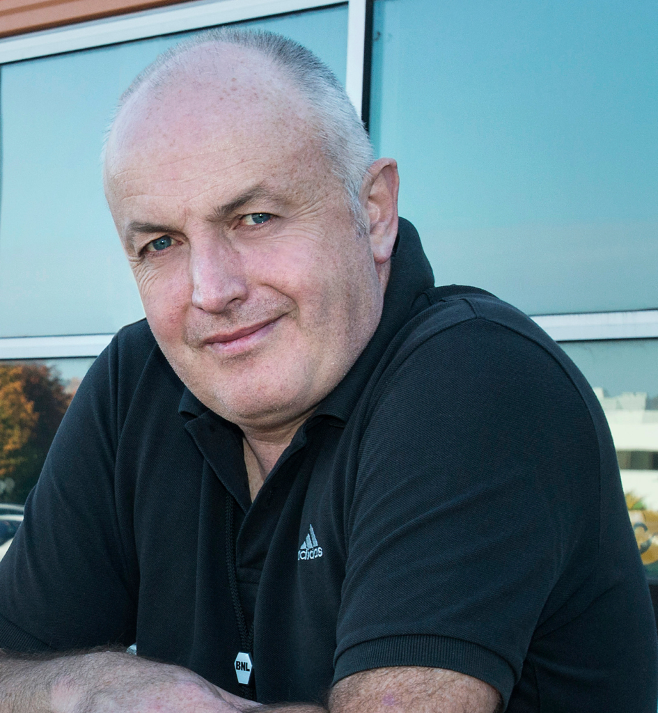
Biography: Sean McSweeney is the delightfully funny and cheerful deputy director of the Photon Division of the NSLS-II at BNL, and the Director of its Center for Biomolecular Structure. Since 1991, he has constructed and operated x-ray beamlines for macromolecular crystallography at synchrotron sites, and has led the way towards automation of synchrotron beamlines. The underlying theme of his research, and that of his group, has been to find methods to make best use of synchrotron light-sources to reveal the complexity of biological systems. His research uses NSLS-II and other infrastructure at BNL for structural biology (of cells and molecules), cryogenic electron microscopy, and using phase-sensitive detection of synchrotron X-rays beams interacting with biological macromolecules to help decipher their structure. Sean got his B.Sc. from the University of Manchester, and went on to obtain his Ph.D in Physical Chemistry, from the same university, in 1991.
